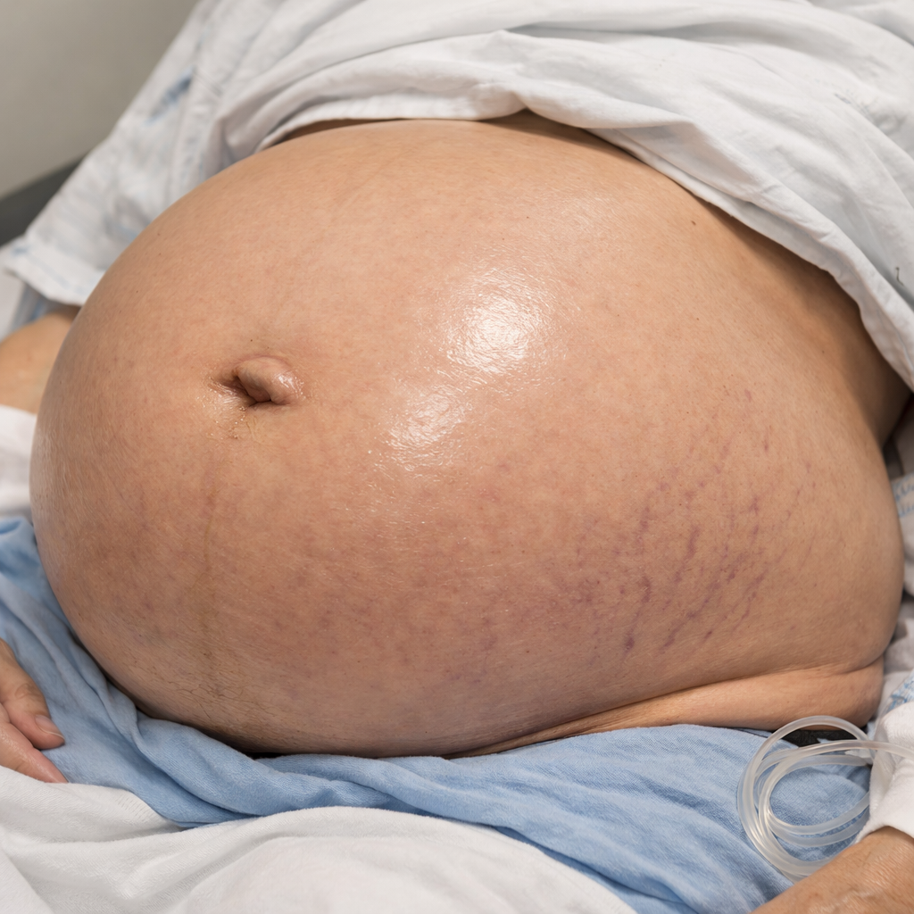
Abdominal swelling is one of those presentations that looks deceptively simple. The patient points to their abdomen and says, “Doctor, it’s getting bigger.” What they are really saying is, something inside me has changed, and I need you to work out what.
The swollen abdomen is not a diagnosis. It is a sign, and like all good signs, it demands interpretation rather than reflex investigation. If you understand the underlying pathophysiology, the abdomen becomes a map rather than a mystery.
Why abdomens swell
All abdominal swellings arise from a surprisingly small number of mechanisms. The abdomen enlarges because something has accumulated, expanded, or escaped its usual boundaries. Fluid may collect silently, as in ascites. Gas may build up noisily, as in bowel obstruction. Solid structures may enlarge or intrude, forming masses that distort the abdominal contour. Fat can redistribute, bowel can load with faeces, and in some cases, life itself grows within the cavity.
The key is not to memorise causes but to recognise behaviour. Fluid shifts when the patient turns. Gas resonates and distends symmetrically. Masses are focal, stubborn, and anatomically loyal. Once you recognise the mechanism, the list of causes narrows dramatically, and your investigations can be purposeful rather than scattergun.
Intestinal obstruction: when movement stops
Few abdominal problems demonstrate pathophysiology as clearly as intestinal obstruction. Here, swelling is the visible consequence of failure of propulsion. Contents accumulate upstream, pressure rises, bowel walls stretch, and physiology begins to unravel.
The cause depends on where the blockage lies. In the small bowel, obstruction is usually a legacy of surgery: adhesions quietly tightening their grip over time. Hernias also play their part, especially when neglected. Tumours and volvulus are less common but no less dangerous. In the large bowel, however, the story changes. In adults, obstruction of the colon is colorectal cancer until proven otherwise, with volvulus and diverticular strictures close behind.
The patient’s symptoms follow predictable rules. Small bowel obstruction declares itself early with vomiting and cramping pain, while the abdomen may still look relatively modest. Large bowel obstruction is slower and more theatrical, producing marked distension, constipation, and late vomiting. When systemic signs appear—tachycardia, fever, rising lactate, the bowel is no longer merely obstructed; it is threatened.
Management begins not with a scalpel but with respect for physiology. Fluids, electrolyte correction, decompression, and careful observation buy time. CT scanning provides clarity. But delay in the face of deterioration costs bowel, and bowel loss costs lives.
Masses in the iliac fossae: anatomy is everything
When swelling localises to the right or left iliac fossa, anatomy takes centre stage. The right iliac fossa is a crowded neighbourhood. Appendix, caecum, terminal ileum, ureter, ovary, and psoas muscle all jostle for space. An inflammatory appendicular mass, a caecal tumour, Crohn’s disease, or gynaecological pathology may all present with similar contours but very different consequences.
The left iliac fossa is quieter but no less dangerous. Here, the sigmoid colon dominates. Diverticular disease is the commonest culprit, forming inflammatory masses or abscesses that mimic malignancy. Faecal loading can deceive the unwary. Cancer lurks quietly and must always be excluded once inflammation settles.
In both regions, imaging is not optional. Ultrasound has its place, particularly in younger patients and pelvic pathology, but CT scanning remains the definitive arbitrator.
Epigastric swellings: the deceptive middle
Swelling in the epigastrium is often misread because it sits at the crossroads of multiple systems. The stomach, pancreas, liver edge, and great vessels all project here, and each leaves a distinctive signature if you know how to look.
A pancreatic pseudocyst emerges weeks after inflammation and feels smooth, fixed, and relentless. A gastric malignancy may present late, hard and unforgiving. A pulsatile epigastric mass is never “just gastritis”; it is an abdominal aortic aneurysm until proven otherwise. The liver, particularly its left lobe, can extend into the epigastrium, moving obediently with respiration and revealing its identity to a careful examiner.
Ultrasound is often the first step, but CT defines the truth.
Imaging: choosing the right window
The investigation of abdominal swelling and pain is as much about restraint as enthusiasm. Plain radiography still has value. An erect chest X-ray revealing free air beneath the diaphragm ends debates instantly. An abdominal X-ray can suggest obstruction or volvulus, but it is a hint, not an answer.
Ultrasound excels at identifying fluid, gallbladder pathology, liver disease, and pelvic causes. It is safe, repeatable, and limited mainly by the operator and the patient’s body habitus.
CT scanning, however, has transformed abdominal medicine. It identifies obstruction, defines masses, reveals inflammation, and exposes complications with ruthless clarity. In the acute abdomen, CT does not replace clinical judgement; it sharpens it.
Contrast studies still have selective roles, particularly in obstruction, where they may diagnose and relieve at the same time, but they are no longer first-line tools.
Bowel obstruction: when swelling turns dangerous
The real danger of bowel obstruction lies not in the swelling itself but in what follows. As pressure builds, venous return is compromised, arterial inflow suffers, and ischaemia develops. Necrotic bowel perforates silently or catastrophically. Bacteria flood the peritoneal cavity. Sepsis follows.
Meanwhile, physiology unravels. Fluid sequestration causes hypovolaemia. Vomiting strips electrolytes and drives metabolic alkalosis. Ischaemia and sepsis drag the patient into acidosis. The abdomen may look unchanged, but the patient is slipping away.
This is why obstruction is never a condition to “watch and wait” casually. Observation is active, vigilant, and brief.
Bringing it all together
Abdominal swelling is a lesson in humility. It reminds us that symptoms are signals, not answers. The abdomen does not lie, but it does require translation. Shape, sound, movement, and behaviour all carry meaning if you know how to interpret them.
Understand the mechanism, localise anatomically, investigate intelligently, and act decisively when danger signals appear. Do this, and the swollen abdomen becomes less of a riddle and more of a story, one that tells you exactly how it wants to end.
Professor’s closing words
The abdomen is generous with clues but ruthless with delay. It will warn you before it collapses, but only once.
Listen carefully.
Think anatomically.
Respect physiology.
Because when the abdomen swells, it is not being dramatic.
It is asking for your attention, urgently, and often for the last time.